A bone spur under foot, often called a heel spur, is one of the most common reasons patients walk into our offices at Achilles Foot and Ankle Center. It’s a small, bony projection that develops along the underside of the heel bone, and while some people never feel it, others deal with sharp, stabbing pain that makes every step miserable.
These growths don’t appear overnight. They form gradually, often over months or years, as the body responds to ongoing stress, inflammation, or tension in the soft tissues of the foot. Understanding what causes them, how to recognize the symptoms, and when to seek professional treatment makes a real difference in how quickly you recover, and whether surgery ever becomes part of the conversation.
Our podiatrists across Central Virginia’s thirteen clinic locations diagnose and treat bone spurs daily, using everything from conservative therapies to advanced surgical techniques when necessary. In this article, we’ll break down exactly what’s happening beneath your foot, why it’s happening, and what your treatment options look like from start to finish.
Why a bone spur forms under the foot
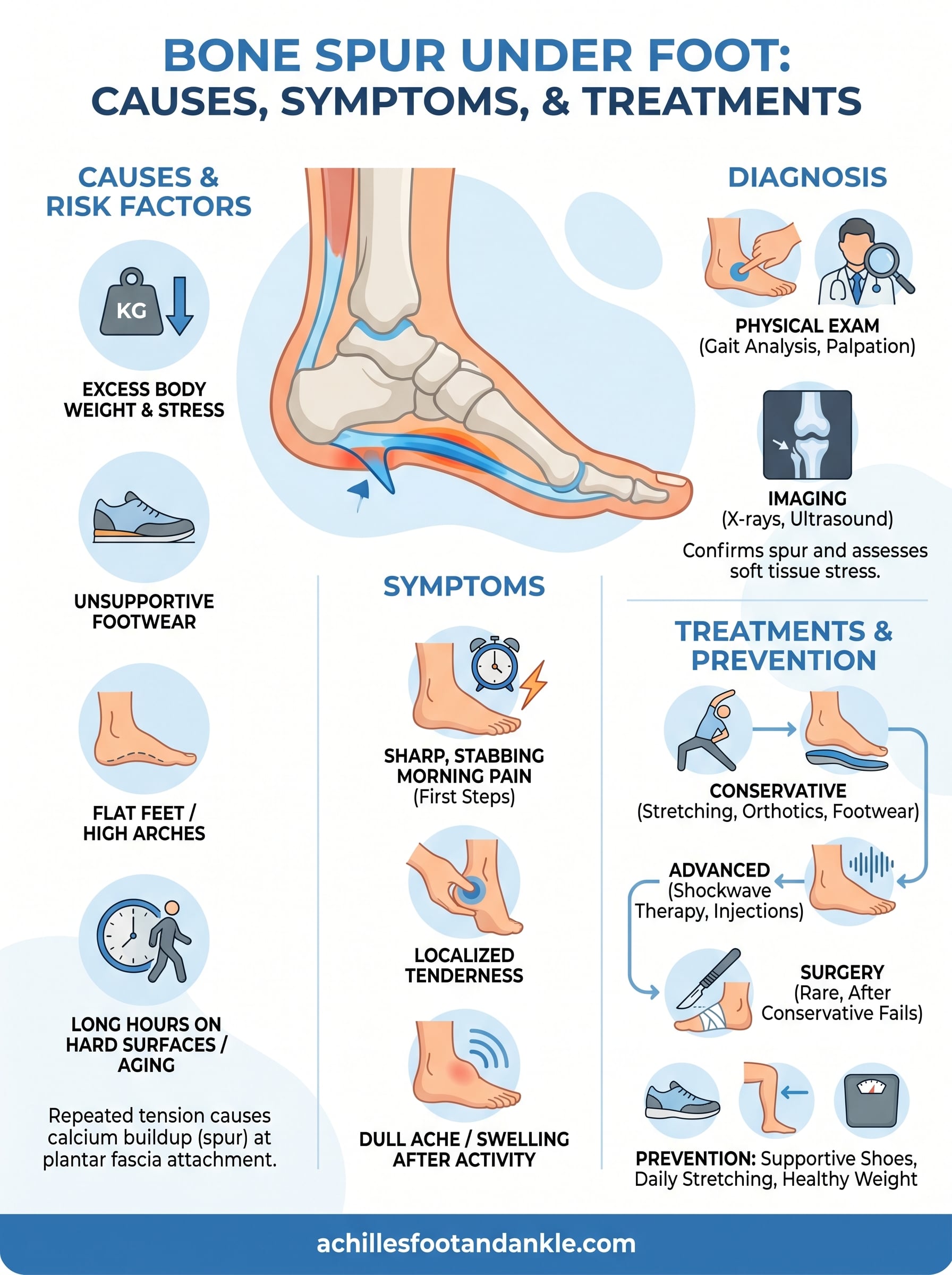
Your body is constantly responding to stress placed on your bones and soft tissues. When the underside of your foot experiences repeated tension or irritation, the heel bone, known as the calcaneus, reacts by depositing extra calcium at the attachment point of the plantar fascia ligament. Over time, that calcium buildup hardens into a bony projection, which is exactly what a bone spur under foot is. The process is slow and often painless at first, which is why many people don’t realize it’s happening until the spur is well established.
The role of plantar fascia tension
The plantar fascia is a thick band of tissue that runs along the bottom of your foot, connecting your heel bone to your toes. Every step you take pulls on this tissue. When that pulling becomes excessive or chronic, tiny microtears form where the fascia attaches to the heel. Your body interprets these microtears as damage and responds by laying down calcium deposits to reinforce the area.
The spur itself is not always the direct cause of pain. The ongoing inflammation in the plantar fascia surrounding it is what usually drives the sharp, stabbing sensation many patients describe.
This is why plantar fasciitis and heel spurs so often show up together on imaging. They are separate conditions, but the same underlying stress typically triggers both. Treating one without addressing the other rarely produces lasting relief.
Lifestyle and biomechanical factors that raise your risk
Certain habits and physical characteristics make you significantly more likely to develop a bone spur. Understanding your personal risk factors gives you a better shot at catching the problem early or preventing it altogether.
The most common contributing factors include:
- Flat feet or high arches: Both alter how weight distributes across your foot, increasing strain on the plantar fascia attachment point
- Excess body weight: More load on your heels means more mechanical stress with every step
- Standing or walking on hard surfaces for long hours: Common in occupations like nursing, retail work, or construction
- Worn-out or unsupportive footwear: Shoes that lack proper arch support accelerate the stress cycle
- Tight calf muscles: Limited ankle flexibility forces the plantar fascia to compensate, pulling harder at the heel
Age also plays a role, since the fat pad protecting your heel naturally thins as you get older, leaving the bone more exposed to impact forces. Combining several of these factors significantly increases the likelihood that a spur will develop and eventually become symptomatic.
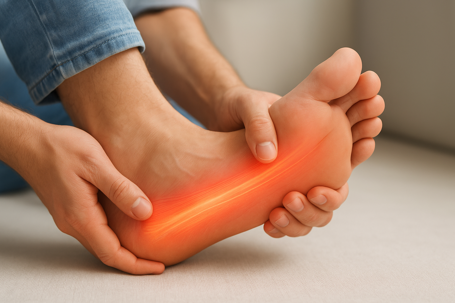
Symptoms and what heel pain really means
Not all heel pain comes from a bone spur under foot, but the pattern is often distinctive enough to raise suspicion quickly. Most patients describe a sharp, stabbing sensation in the heel that hits hardest with the first few steps in the morning or after sitting for a long stretch. Once you’ve been moving for a few minutes, the pain may ease, only to return after extended standing or walking.
The classic morning pain pattern
That first-step pain has a specific explanation. When you rest, the plantar fascia contracts and partially heals in a shortened position. The moment you put weight on your foot, you stretch that tight tissue abruptly, pulling against the inflamed attachment point near the spur. Patients frequently describe it as stepping on a sharp pebble or nail, even when nothing is there.
This pattern of pain that improves briefly with movement but worsens again after prolonged activity is one of the clearest early indicators that your plantar fascia and heel are under significant stress.
Other symptoms you should not ignore
Beyond morning pain, you may notice localized tenderness when pressing directly on the bottom of your heel. Some people experience swelling around the heel area, particularly after a long day on their feet. A persistent dull ache that spreads toward the arch can also develop as the condition progresses.
If the pain starts changing how you walk, causing you to shift weight to the outer edge of your foot or shorten your stride, that compensation pattern creates secondary problems in your knees, hips, or lower back. Addressing the heel issue before those downstream effects develop is the reason early evaluation matters.
How podiatrists diagnose a bone spur
When you come in with heel pain, a podiatrist does not jump straight to imaging. The diagnostic process starts with a thorough conversation about your symptoms, including how long the pain has been present, when it’s worst, and what activities or footwear trigger it. That history alone often points clearly toward a bone spur under foot before any test is ordered.
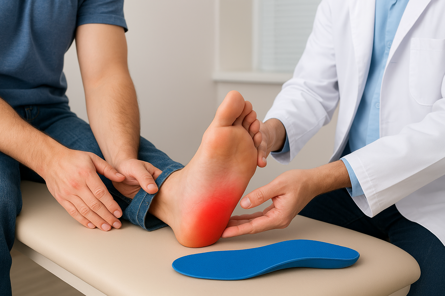
Physical examination
Your podiatrist will press directly on the bottom of your heel to locate the exact spot causing pain. This manual palpation helps distinguish a heel spur from other conditions like Achilles tendinitis or tarsal tunnel syndrome, which produce pain in different locations. They will also assess your range of motion, arch shape, and gait pattern to identify biomechanical issues contributing to the problem.
A thorough physical exam often reveals just as much useful information as imaging, especially when the pain pattern and tender point match the classic presentation of plantar fascia stress.
Gait analysis also plays a key role. Watching you walk reveals how your foot strikes the ground and whether your arch collapses inward with each step. Those movement patterns directly influence where stress concentrates on your heel bone, and correcting them is often central to treatment.
Imaging to confirm the diagnosis
X-rays are the standard first step for confirming a bone spur. A spur typically appears as a pointed, hook-shaped projection on the underside of the calcaneus, and it can range from a few millimeters to over a centimeter in length. If soft tissue involvement needs closer evaluation, your podiatrist may order diagnostic ultrasound or an MRI to assess the plantar fascia directly and rule out partial tears before deciding on a treatment plan.
How to treat a bone spur under the foot
Treatment for a bone spur under foot almost always starts with conservative methods, and the majority of patients recover fully without ever needing surgery. Your podiatrist will build a plan around your specific symptoms, biomechanical factors, and how long the condition has been present. Starting treatment early gives those conservative approaches the best chance of working.
Conservative treatments that work first
The first line of care focuses on reducing inflammation and relieving pressure at the heel. This typically includes a combination of physical therapy to stretch the plantar fascia and calf muscles, custom orthotics to redistribute load more evenly across your foot, and anti-inflammatory medications or corticosteroid injections to calm the inflamed tissue around the spur. Switching to supportive footwear with adequate cushioning also removes a significant amount of daily stress from your heel.
Most patients see meaningful improvement within six to twelve weeks when they consistently follow a conservative treatment plan that addresses both the symptoms and the underlying biomechanical cause.
For cases that do not respond to initial therapy, your podiatrist may recommend extracorporeal shockwave therapy (ESWT), a non-invasive procedure that delivers targeted sound waves to stimulate healing in the affected tissue. This option works well for patients with persistent plantar fascia pain who want to avoid an incision entirely.
When surgery becomes the right option
Surgery is reserved for the small percentage of patients whose pain does not improve after six to twelve months of dedicated conservative treatment. The procedure typically involves releasing part of the plantar fascia and removing the spur itself. Recovery requires several weeks of limited weight bearing, but outcomes are generally strong for patients who have exhausted non-surgical options and are committed to post-operative rehabilitation.
Prevention and when to see a podiatrist
Preventing a bone spur under foot comes down to reducing the repetitive stress that triggers calcium deposits in the first place. Small, consistent habits protect your heel bone over time and significantly lower your risk of developing both spurs and the plantar fascia inflammation that typically accompanies them.
Steps you can take to protect your heels
Wearing supportive footwear is the single most effective preventive step most people overlook. Shoes with adequate arch support, cushioned heel cups, and proper fit reduce the load your plantar fascia absorbs with every step. Replacing worn shoes before the midsole breaks down keeps that protection consistent.
Stretching your calf muscles and plantar fascia daily also makes a significant difference. Tight calves pull on the heel attachment point throughout the day, and a few minutes of targeted stretching in the morning and before activity reduces that tension considerably. Maintaining a healthy body weight directly lowers the mechanical force your heels absorb with every step you take.
Signs it is time to call a podiatrist
You should contact a podiatrist when heel pain persists for more than a week or two, when morning stiffness is not improving, or when discomfort starts changing how you walk. Waiting too long allows compensatory movement patterns to develop, which creates additional strain on your knees, hips, and lower back.
Catching heel pain early gives conservative treatments the best chance to resolve the problem without injections or surgery.
Pain that is sharp, constant, or worsening despite rest and basic home care is a clear signal that professional evaluation is overdue. A podiatrist can identify whether a spur is present, assess your biomechanics and gait, and get you started on a targeted plan before the condition becomes harder to treat.
Next steps
A bone spur under foot is a manageable condition, and the path forward is straightforward once you know what you’re dealing with. Most patients recover fully with conservative care, particularly when they start treatment before the pain becomes severe or starts affecting how they move. The key is acting on the warning signs rather than waiting them out.
If heel pain is slowing you down, interfering with your mornings, or shifting the way you walk, that is your signal to get a professional evaluation. Our podiatrists at Achilles Foot and Ankle Center work across thirteen locations in Central Virginia and offer same-day appointments for patients who need answers quickly. You do not need to guess about what is happening in your heel or manage the pain on your own. Schedule a same-day appointment and get a clear diagnosis and a treatment plan built specifically around your foot.