That nagging pain on the top of your foot or the back of your heel that sharpens every time you take a step, it might not be "just" inflammation. Foot bone spur symptoms often develop gradually, and many people walk around with bony growths (called osteophytes) for months before connecting their discomfort to an actual structural change in the bone. The tricky part is that not all bone spurs cause pain, which means some go unnoticed while others make even basic movement feel miserable.
Understanding what to look for matters. A visible bump, stiffness in a joint, or swelling that doesn’t resolve with rest can all point toward a bone spur, but these signs also overlap with other foot conditions. Knowing the difference helps you avoid guessing and get the right care sooner rather than later. At Achilles Foot and Ankle Center, our podiatry team across Central Virginia diagnoses and treats bone spurs regularly, using advanced imaging and both surgical and non-surgical approaches tailored to each patient.
This article breaks down the specific symptoms of foot bone spurs, what causes them to form, and the care options available to get you back on your feet without pain.
What a foot bone spur is and where it forms
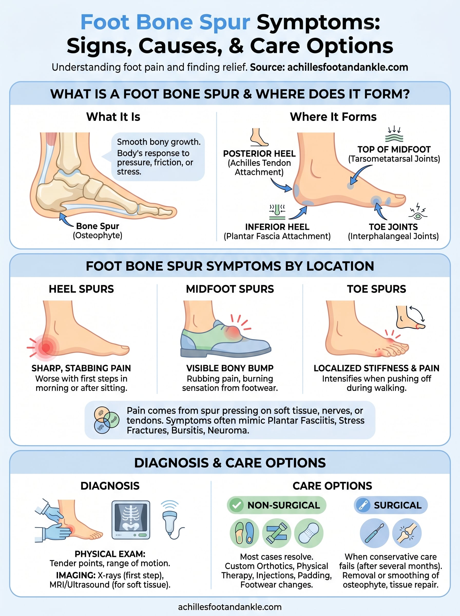
A bone spur, or osteophyte, is a smooth bony projection that grows along the edge of an existing bone. Your body forms these deposits as a direct response to prolonged pressure, friction, or mechanical stress on a specific area. The process is slow and gradual, often taking months or years before it produces noticeable changes. Over time, calcium accumulates at a stress point, and your body lays down extra bone tissue to reinforce that area. Bone spurs are not automatically dangerous, but when they press against nearby soft tissue, nerves, or tendons, they generate the pain and stiffness most people recognize as foot bone spur symptoms.
How bone spurs develop in the foot
The foot absorbs tremendous mechanical load with every step you take, making it one of the most common sites for osteophyte formation. Repetitive stress from walking, running, or standing for extended periods creates micro-trauma at bone surfaces, particularly where tendons and ligaments attach to bone. Your body responds to this ongoing irritation by depositing extra calcium at the attachment site, a process called enthesopathy. Conditions like plantar fasciitis, Achilles tendinopathy, and osteoarthritis accelerate this process because they keep the attachment site in a persistent state of low-grade inflammation, giving osteophytes a favorable environment to grow.
Most bone spurs develop over several years of accumulated stress at a specific location in the foot, which is why many patients are surprised to learn a spur exists when they first see an X-ray.
Where bone spurs most commonly form
The location of a spur largely determines which symptoms you experience and how severe they become. Not every part of the foot carries the same level of mechanical stress, so osteophytes tend to concentrate in predictable areas. The inferior heel is the single most common site, specifically where the plantar fascia attaches to the calcaneus bone. You can also develop spurs at the back of the heel where the Achilles tendon inserts, along the top of the midfoot at the tarsometatarsal joints, and at the small joints of the toes.
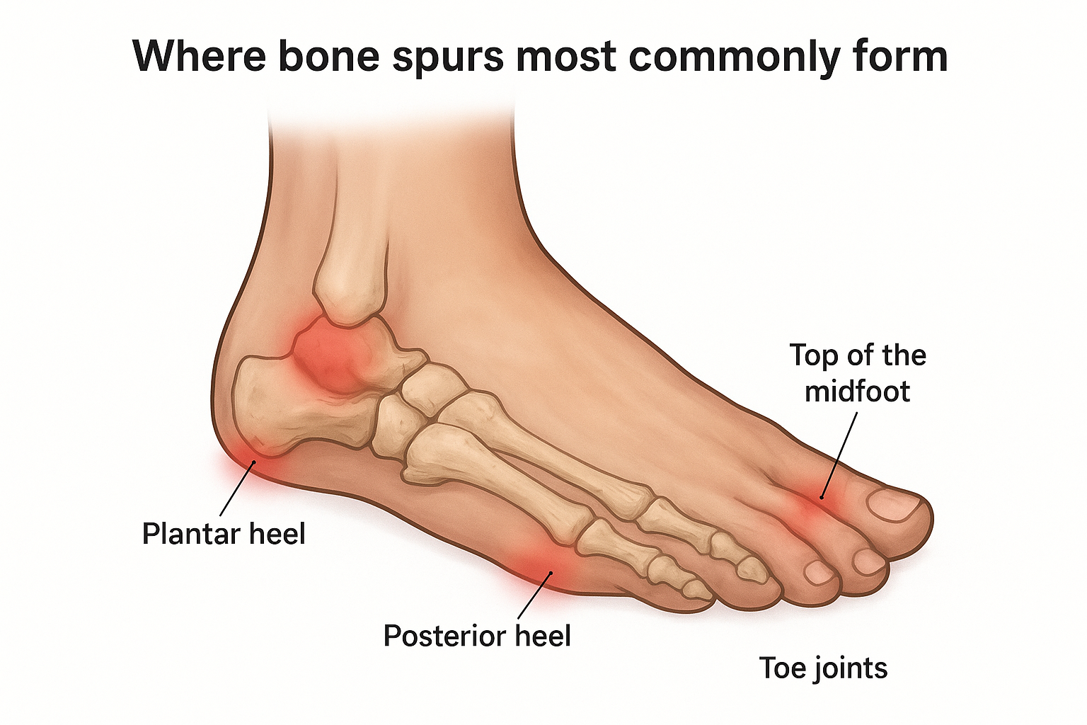
Common formation sites include:
- Plantar heel (inferior calcaneus): beneath the heel, closely associated with plantar fasciitis
- Posterior heel: at the Achilles tendon insertion, sometimes called a Haglund deformity
- Top of the midfoot: over the navicular or cuneiform bones, often irritated by tight footwear
- Toe joints: along interphalangeal joints, frequently linked to hammertoes or arthritis
- Ball of the foot: near the metatarsal heads, commonly from repeated impact during activity
Foot bone spur symptoms by location
Foot bone spur symptoms vary depending on exactly where the osteophyte has formed. The same type of bony growth can cause completely different sensations depending on which structures it presses against, which is why two people with bone spurs can describe their pain in entirely different ways.
Heel bone spurs
Heel spurs on the bottom of the foot typically produce a sharp, stabbing pain that hits hardest when you first stand up in the morning or after sitting for a long stretch. That first step out of bed can feel like stepping on a small stone. Posterior heel spurs, located where the Achilles tendon attaches, tend to cause a different pattern: a deep ache or swelling at the back of the heel that worsens with activity and makes the heel sensitive to the pressure of a shoe collar.
Pain that eases after a few minutes of walking but returns sharply after extended rest is a strong indicator of an inferior heel spur pressing against the plantar fascia.
Midfoot and toe spurs
Spurs along the top of the midfoot often announce themselves as a visible, bony bump that rubs painfully against the upper part of your shoe. You may notice redness, a burning sensation, or even numbness across the top of the foot if the growth presses on a small nerve branch. Toe joint spurs, particularly near the big toe or smaller interphalangeal joints, produce localized stiffness, reduced range of motion, and pain that intensifies when you push off during walking or climbing stairs.
Why foot bone spurs cause symptoms and what they mimic
A bone spur on its own is a smooth, rounded structure, and it doesn’t produce pain simply by existing. Symptoms emerge when the osteophyte presses against surrounding soft tissue, compressing tendons, nerves, bursae (small fluid-filled sacs), or joint cartilage. Your body responds to this compression with inflammation, which is what you actually feel as pain, swelling, and stiffness. The more load your foot absorbs during daily activity, the more that inflammation cycles and the worse your discomfort becomes.
Pain from a bone spur is almost always secondary, meaning the spur triggers a tissue response, and that response is what you feel.
Why the spur itself isn’t always the direct cause of pain
Many people have visible spurs on X-ray but report no foot bone spur symptoms at all, because the osteophyte hasn’t grown into a position where it contacts sensitive tissue. When a spur does press against a nerve, it can cause burning, tingling, or numbness instead of straightforward mechanical pain. The precise angle and size of the growth determines which nearby structures take the pressure and how intense your symptoms become over time.
Conditions that mimic bone spur symptoms
Several foot conditions produce overlapping symptoms, which makes self-diagnosis unreliable without imaging. Plantar fasciitis, stress fractures, bursitis, and Morton’s neuroma can all cause heel or forefoot pain that feels identical to a spur from the inside. Peripheral neuropathy, particularly in patients with diabetes, adds another layer of complexity because it generates burning and numbness that closely mirrors nerve compression from an osteophyte. Getting an accurate diagnosis requires professional evaluation rather than guesswork based on symptoms alone.
How doctors diagnose foot bone spurs
Diagnosing a bone spur starts with a conversation. Your doctor will ask about the location, timing, and pattern of your symptoms, including whether your pain spikes in the morning, worsens with specific footwear, or follows a period of increased activity. This history helps narrow down which structure is likely involved before any imaging takes place. Reporting your foot bone spur symptoms accurately gives your provider a clearer starting point and reduces the chance of missing an overlapping condition.
Physical examination
During the physical exam, your podiatrist will press along specific landmarks on your foot to identify tender points and compare them against known attachment sites for tendons and ligaments. You may be asked to flex or extend your foot against resistance so the clinician can assess how load-bearing movement affects your discomfort. Joint range of motion, visible deformity, and localized swelling all factor into whether a spur is suspected before imaging confirms it.
A tender spot directly at the base of the heel or at the Achilles insertion, combined with pain on first steps, gives a podiatrist strong clinical grounds to suspect an osteophyte.
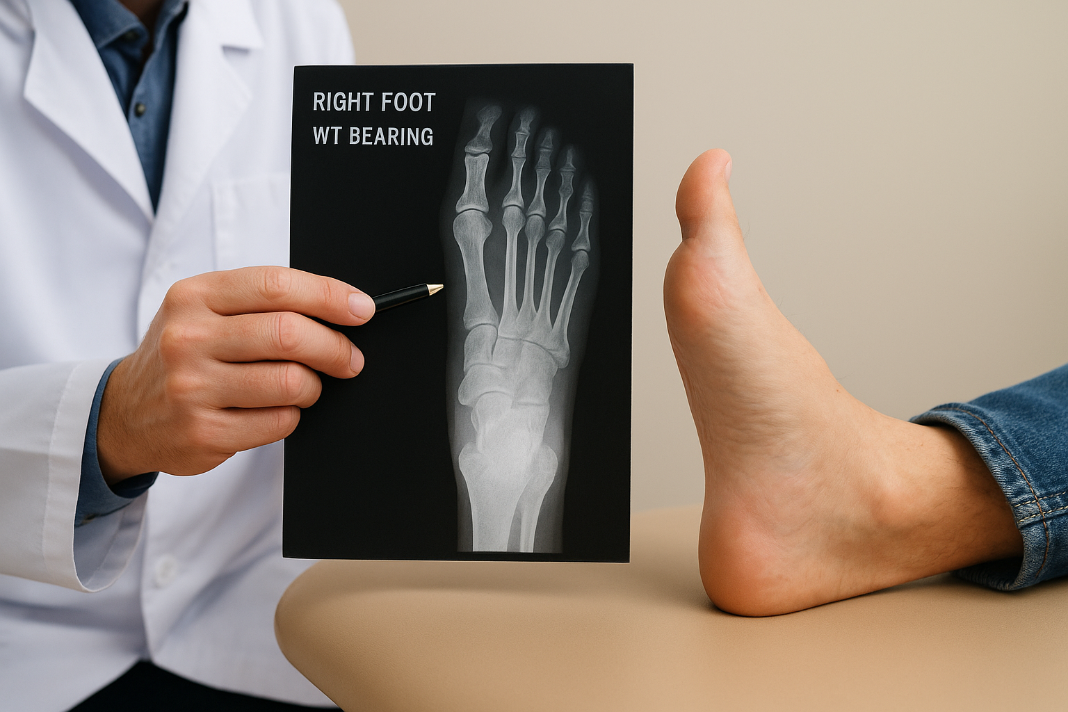
Imaging tests
Your doctor will typically order weight-bearing X-rays as the first imaging step because they capture the foot under realistic load and reveal calcified deposits at bone surfaces with good clarity. X-rays confirm the presence, size, and location of a spur efficiently. In cases where soft tissue involvement needs closer evaluation, such as nerve compression or tendon damage, an MRI or diagnostic ultrasound provides detail that X-rays cannot, showing how the osteophyte interacts with surrounding structures.
Care options for painful foot bone spurs
Treatment focuses on relieving the inflammation and soft tissue irritation that the spur triggers, not always on removing the growth itself. Many patients manage their foot bone spur symptoms successfully without surgery, especially when they address the condition before it progresses. Your treatment plan depends on which location is affected, how long symptoms have been present, and how much the pain limits your daily activity.
Non-surgical approaches
Conservative care resolves the majority of painful bone spur cases. Custom orthotics redistribute pressure away from the spur, reducing the mechanical load that feeds the cycle of irritation. Physical therapy strengthens the surrounding muscles and improves flexibility in the tendons that attach near the osteophyte. Corticosteroid injections, administered under ultrasound guidance, reduce acute inflammation when physical therapy alone isn’t enough to get you moving comfortably again. Padding, activity modification, and switching to footwear with a wider toe box or deeper heel cup can also significantly lower the daily friction the spur generates.
Most patients see meaningful improvement within six to twelve weeks of consistent conservative treatment when they address the root cause alongside the spur itself.
When surgery makes sense
Surgery becomes the right option when conservative treatment fails to reduce your symptoms after several months of dedicated effort, or when imaging shows the spur is compressing a nerve or tendon in a way that cannot be addressed with external measures. Your surgeon removes or smooths the osteophyte and, when necessary, repairs the damaged tissue around it. Minimally invasive techniques allow for smaller incisions, faster recovery, and less post-operative discomfort compared to traditional open procedures.
Next steps if you suspect a bone spur
If your foot bone spur symptoms match what you’ve read here, the smartest move is to stop guessing and get a proper evaluation. A podiatrist can confirm whether a spur is present, identify its exact location, and determine which structures it affects before your pain has a chance to worsen. Waiting typically makes treatment harder, since prolonged inflammation can damage the tendons and soft tissue surrounding the osteophyte.
Treatment works best when it starts early. Most people respond well to conservative care and never need surgery, but that outcome depends on catching the condition at a manageable stage. Achilles Foot and Ankle Center offers same-day appointments across thirteen Central Virginia locations, so you don’t have to sit with foot pain while waiting weeks for answers. Schedule a same-day appointment today and get a clear picture of what’s actually happening in your foot.