A bone spur in your foot often starts without any noticeable symptoms, just a small, bony projection forming along a joint or where a tendon connects to bone. But over time, that growth can press against soft tissue, nerves, or other bones, turning every step into a painful reminder that something isn’t right. Understanding bone spur foot causes helps you recognize whether you’re at risk and, more importantly, what you can do about it before the problem gets worse. These bony growths don’t appear randomly. They develop as your body’s response to pressure, friction, or damage happening at specific points in the foot.
Several factors drive bone spur formation, from the way you walk and the shoes you wear to underlying conditions like arthritis, plantar fasciitis, and obesity. Some people are simply more prone to developing them based on foot structure or age-related joint changes, while others push their feet past a breaking point through repetitive stress from work or athletics. At Achilles Foot and Ankle Center, our podiatrists across Central Virginia diagnose and treat bone spurs regularly, using everything from digital imaging to pinpoint the growth to conservative and surgical options tailored to each patient’s situation.
This article breaks down exactly why bone spurs form in the foot, which risk factors matter most, and how to recognize the signs early. Whether you’re dealing with heel pain that won’t quit or you’ve just been told you have a spur on an X-ray, you’ll walk away with a clear picture of what’s happening and what steps to take next.
How bone spurs form in the foot
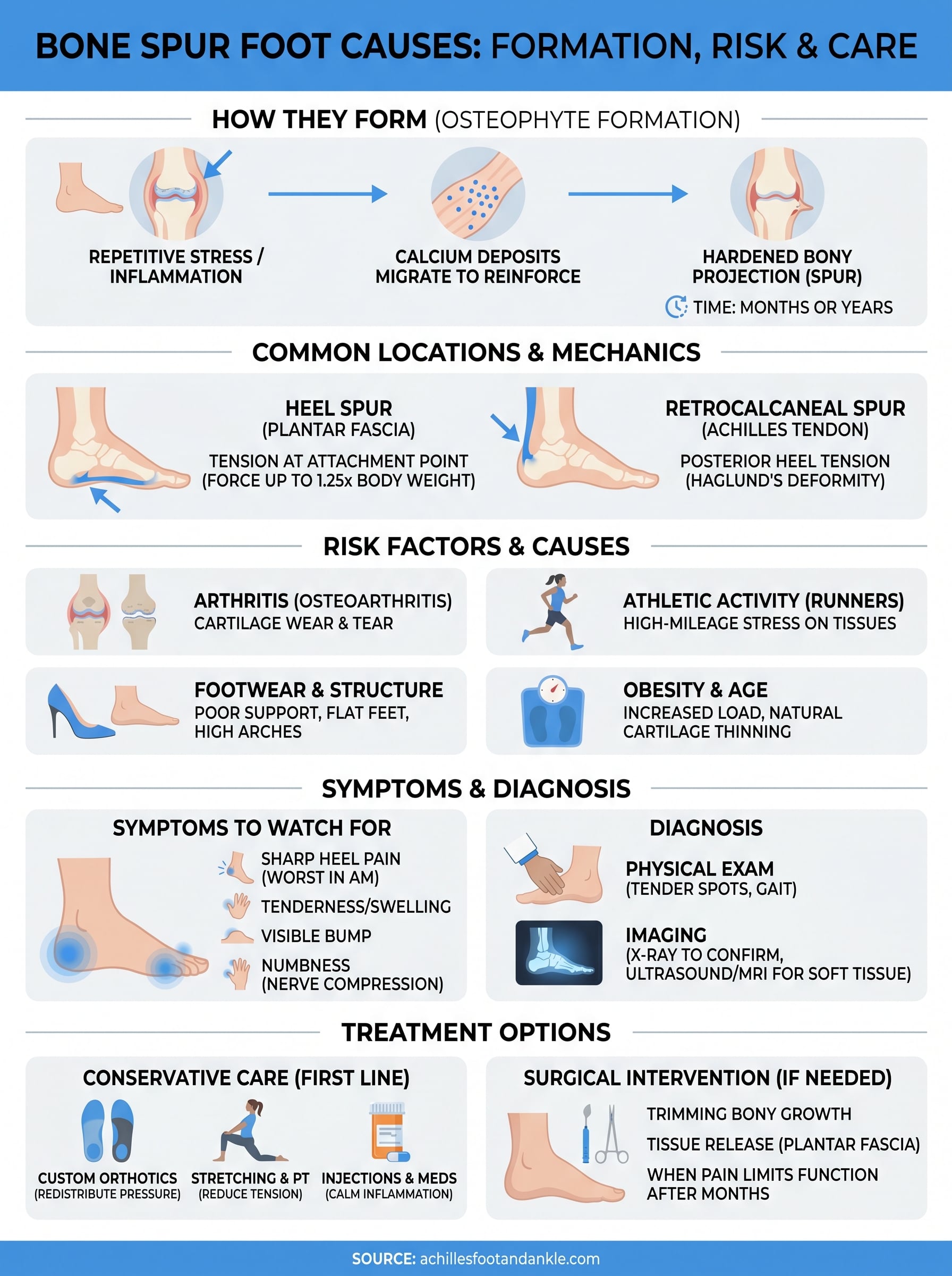
Your body builds bone spurs through a process called osteophyte formation, and it starts as a protective response. When soft tissue around a joint or bone insertion point experiences repeated stress or damage, your body interprets that stress as a threat and begins depositing extra calcium at the site. Over time, those calcium deposits harden into a bony projection. The process isn’t sudden; it typically develops over months or even years before it becomes symptomatic.
The role of inflammation and calcium deposits
Inflammation is the trigger that sets this process in motion. When ligaments, tendons, or joint cartilage get irritated repeatedly, the surrounding tissue signals the body to reinforce that area. Calcium migrates to the inflamed zone and gradually solidifies into a hard growth. This is your body attempting to stabilize a vulnerable spot, but the result can press on nerves, bursa sacs, or neighboring tendons. Understanding this mechanism is central to understanding bone spur foot causes, because it shows why addressing inflammation early can sometimes prevent the spur from growing larger.
Bone spurs are not a disease in themselves; they are a sign that your foot has been under chronic stress for a long time.
The location of inflammation determines exactly where the spur forms. If the plantar fascia pulls repeatedly at its heel attachment, a heel spur develops at the bottom of the calcaneus. If cartilage in a toe joint breaks down, spurs form along the joint margins instead. The mechanical stress driving that inflammation varies from person to person, which is why two people with identical activity levels can end up with very different outcomes.
Why the heel is the most common site
Your heel absorbs a tremendous amount of force with every step you take. On average, it handles forces equivalent to 1.25 times your body weight during normal walking, and that figure climbs sharply during running or jumping. The plantar fascia attaches directly to the heel bone, and when that ligament is under repeated tension, the attachment point becomes the ideal environment for spur formation. That’s why heel spurs so often appear alongside plantar fasciitis, a condition driven by exactly this kind of chronic pulling stress on the fascia.
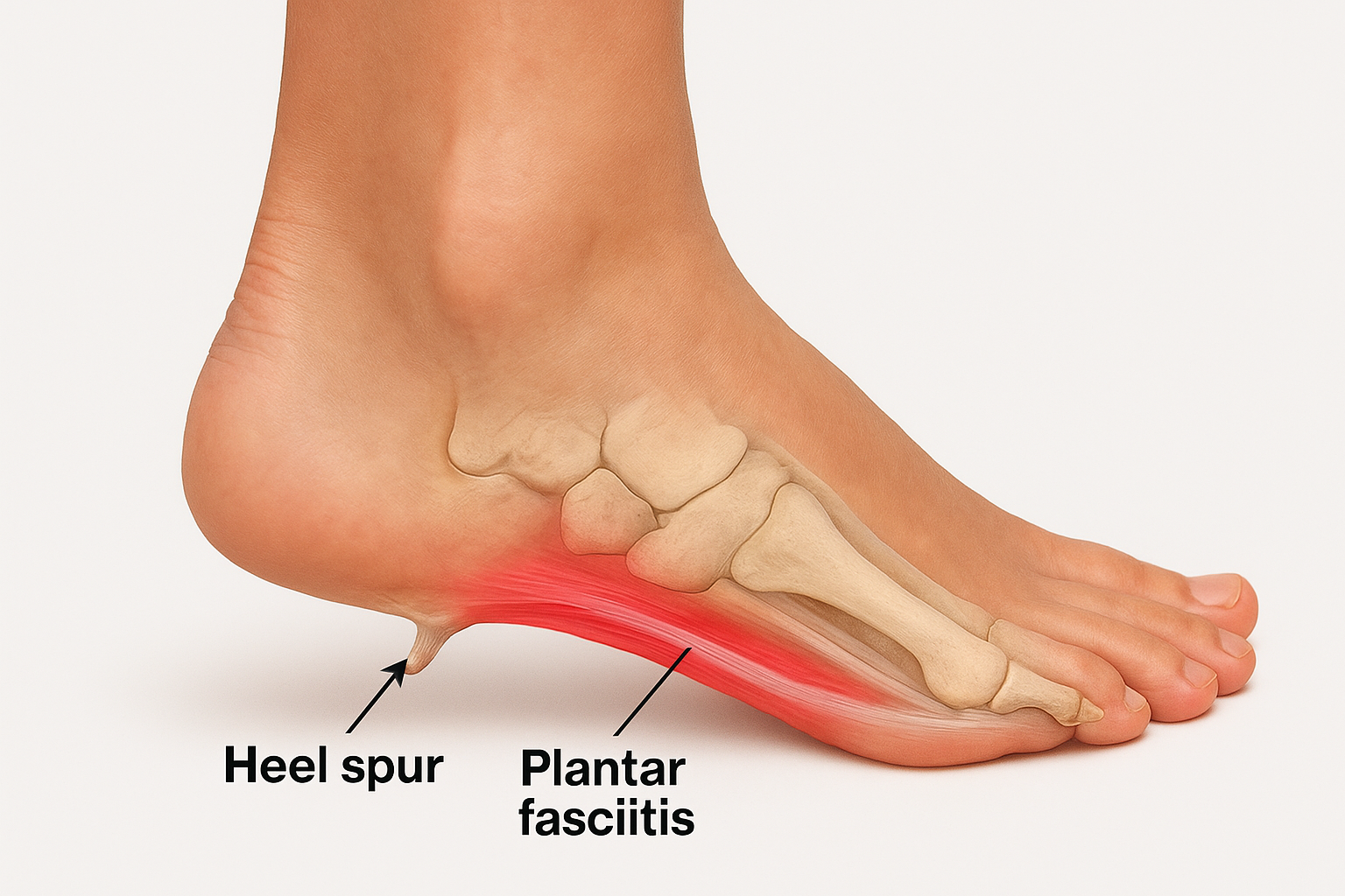
At the back of the heel, the Achilles tendon attaches directly to the calcaneus, creating another common spur location. This type, often called a Haglund’s deformity or retrocalcaneal spur, develops through similar mechanics but affects the posterior heel rather than the bottom, producing pain and swelling where the tendon meets the bone.
Bone spur foot causes and who’s at risk
Several distinct factors contribute to bone spur formation, and knowing which ones apply to you can make a real difference in how you approach prevention or treatment. Repetitive mechanical stress is the most consistent thread running through all bone spur foot causes, whether that stress comes from athletic activity, occupational demands, or structural issues in the foot itself.
Conditions and lifestyle factors that drive spur growth
Arthritis is one of the leading contributors, particularly osteoarthritis, which gradually wears down joint cartilage and prompts the body to produce bony growths at joint margins as a stabilizing response. Plantar fasciitis, tight calf muscles, flat feet, and high arches all create abnormal tension patterns that increase the likelihood of heel spurs. Obesity adds another layer of risk by multiplying the compressive forces your feet handle with every step.
Common conditions linked to bone spur development include:
- Osteoarthritis and rheumatoid arthritis
- Plantar fasciitis and Achilles tendinopathy
- Flat feet, high arches, and other structural imbalances
- Diabetes-related foot changes
Wearing shoes with poor arch support or a tight toe box over many years is one of the most preventable contributors to bone spur development.
Who faces the highest risk
Certain groups consistently show up at higher rates in bone spur cases. Runners and long-distance athletes log high-mileage weeks that place sustained stress on plantar fascia and Achilles tendon attachment points. People over 40 also face increased risk because cartilage naturally thins with age, making joint surfaces more vulnerable to the friction that triggers calcium deposits.
Additional high-risk profiles include:
- Workers who spend long shifts standing on hard surfaces
- Individuals with a family history of arthritis or a prior foot injury
- People who are significantly overweight, placing chronic excess load on foot structures
Symptoms and what foot pain can mean
Bone spurs don’t always cause pain. Some people discover them only when a doctor orders an X-ray for an unrelated issue. But when a spur presses against surrounding soft tissue or nerves, the symptoms can range from a dull ache to sharp, stabbing pain that changes how you walk. Recognizing what your foot is telling you early gives you a much better chance of managing the problem before it limits your daily activity.
Common symptoms to watch for
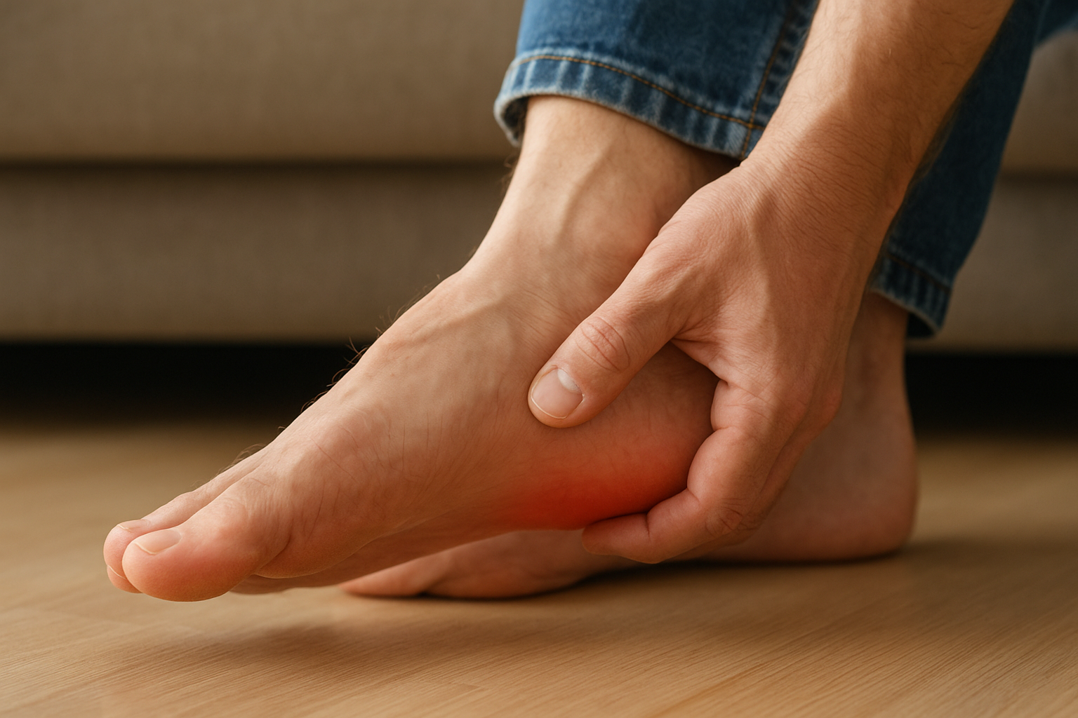
The symptom pattern you experience often points directly to the location and size of the spur. Heel spurs typically produce a sharp pain at the bottom of the heel that feels worst with your first steps in the morning or after sitting for a long period. That pain tends to ease as you move but returns after extended activity. Spurs along toe joints create a more localized ache, visible swelling, or a sensation of grinding when you bend the toe.
Symptoms you might notice include:
- Sharp heel pain with the first steps after rest
- Tenderness or swelling along the arch, heel, or toe joints
- A visible bump or hardened lump on the foot
- Numbness or tingling if the spur compresses a nearby nerve
- Difficulty wearing certain shoes due to pressure on a specific spot
When symptoms signal something serious
If your foot pain is persistent, worsening, or accompanied by swelling and numbness, that combination goes beyond normal soreness. These signs often mean a spur is actively irritating a nerve or tendon, and waiting typically makes the underlying bone spur foot causes harder to treat effectively.
Pain that forces you to alter your gait is a signal worth taking seriously, because compensating for one sore spot often creates new problems in the knee, hip, or lower back.
How doctors diagnose foot bone spurs
Diagnosing a bone spur starts with a conversation. Your podiatrist will ask about your pain history, activity level, and footwear habits to build a picture of what’s been stressing your foot. This background information often points directly to the most likely bone spur foot causes before any imaging is ordered, allowing the doctor to focus the physical examination on the most relevant areas.
Physical examination and what the doctor checks
During the physical exam, your doctor will press along the heel, arch, and toe joints to identify tender spots and any palpable bony prominences. They’ll also assess your range of motion and watch how you walk. Gait abnormalities like overpronation or an uneven push-off pattern are strong indicators that a specific part of the foot has been absorbing excessive stress, which directly informs where a spur is likely developing.
A thorough physical exam often narrows the diagnosis significantly before any imaging is needed, saving time and reducing unnecessary testing.
Imaging and confirming the diagnosis
X-rays are the standard tool for confirming a bone spur. They provide a clear view of bony structures and show the spur’s exact size and location. Your doctor can use this image to determine how much space the spur occupies and whether it’s pressing against a neighboring structure. When soft tissue involvement is suspected, ultrasound or MRI provides additional detail about the condition of surrounding tendons, ligaments, and bursa sacs that an X-ray alone cannot capture. Together, these tools give your care team everything needed to design an effective treatment plan.
Treatment options and what helps
Treatment works best when it matches both the severity of your symptoms and the underlying bone spur foot causes driving the problem. Most cases respond well to conservative care, meaning surgery is rarely the first step. Your podiatrist will typically start with the least invasive options and adjust based on how your foot responds over the following weeks.
Conservative treatments first
Non-surgical approaches address the inflammation and mechanical stress that allow a spur to keep causing pain. Custom orthotics redistribute pressure away from the spur site, which often provides significant relief during daily activity. Stretching programs targeting the plantar fascia and calf muscles reduce the pulling tension at the heel. Corticosteroid injections, physical therapy, and anti-inflammatory medication round out the conservative toolkit and work together to calm the tissue irritation surrounding the spur.
Switching to supportive footwear with adequate arch support and cushioning is one of the simplest changes that can reduce bone spur symptoms within days.
Common non-surgical options your doctor may recommend:
- Custom orthotics or prefabricated arch support inserts
- Targeted stretching and physical therapy exercises
- Corticosteroid or ultrasound-guided injections
- Night splints to reduce morning heel pain
- Activity modification and weight management
When surgery becomes necessary
If conservative treatment fails after several months and your pain continues to limit normal function, surgical removal of the spur becomes a viable option. The procedure involves trimming the bony growth and, where applicable, releasing tight associated tissue like the plantar fascia. Recovery time varies depending on the location of the spur and the complexity of the procedure, but most patients return to normal activity within a few weeks following minimally invasive techniques.
What to do next
Bone spurs don’t get smaller on their own, and the underlying bone spur foot causes rarely resolve without some form of targeted intervention. If your foot pain has lasted more than a few weeks, changed how you walk, or started limiting activities you enjoy, that’s your cue to get a professional opinion rather than wait it out. Early diagnosis means more treatment options and a faster path back to pain-free movement.
Your first step is a thorough evaluation from a podiatrist who can confirm whether a spur is present, identify what’s driving it, and build a treatment plan that fits your life. At Achilles Foot and Ankle Center, our team across Central Virginia uses digital imaging and hands-on examination to give you a clear answer quickly. You don’t need to keep guessing about what’s causing your heel or foot pain. Schedule a same-day appointment and get the answers you need today.