That sharp, stabbing pain under your heel when you step out of bed in the morning, it could be a bone spur on bottom of foot, a bony calcium deposit that develops along the underside of your heel bone. Millions of Americans deal with this condition, and while the spur itself isn’t always the direct source of pain, the inflammation and soft tissue irritation surrounding it can make even simple activities like walking feel miserable.
Heel spurs rarely appear out of nowhere. They typically develop over months or years in response to repeated stress, and they’re closely tied to plantar fasciitis, the most common cause of heel pain. Understanding what’s actually happening beneath your skin is the first step toward getting relief and preventing the problem from getting worse.
At Achilles Foot and Ankle Center, our podiatrists diagnose and treat heel spurs across our Central Virginia locations every day, using everything from conservative therapies to advanced surgical options when needed. In this article, we’ll walk you through what bone spurs on the bottom of the foot look like, what causes them, how they’re diagnosed, and the full range of treatment options available to get you back on your feet without pain.
What a bone spur on the bottom of the foot is
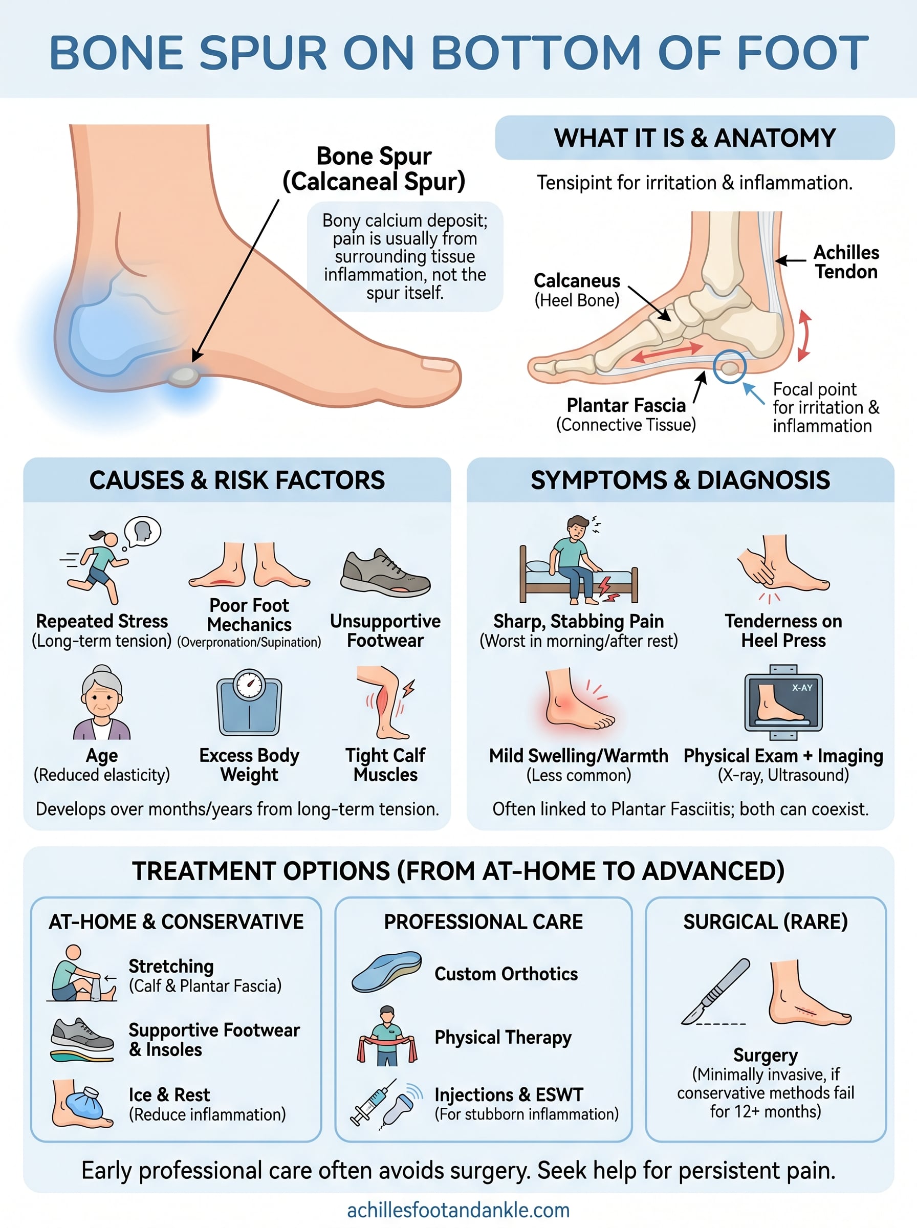
A bone spur, also called a calcaneal spur when it forms on the heel, is a bony projection that grows on the underside of your heel bone (the calcaneus). Your body builds this extra calcium deposit in response to long-term stress, friction, or tension on the bone. The spur itself is not a sharp spike cutting into your tissue. It is a smooth, rounded extension of bone that forms gradually over months or years. Many people carry a bone spur on bottom of foot without ever knowing it because the spur causes no direct pain on its own.
The pain most people attribute to a heel spur is usually coming from the inflamed soft tissue surrounding it, not the bony growth itself.
The anatomy behind the spur
Your heel bone sits at the base of your foot and absorbs an enormous amount of force with every step. When the soft tissues and tendons attached to the bottom of the heel are under repeated or prolonged stress, your body responds by depositing calcium at the site where that tension is greatest. Over time, those deposits accumulate and harden into a bony outgrowth that extends forward along the sole of the foot, sometimes reaching up to half an inch in length.
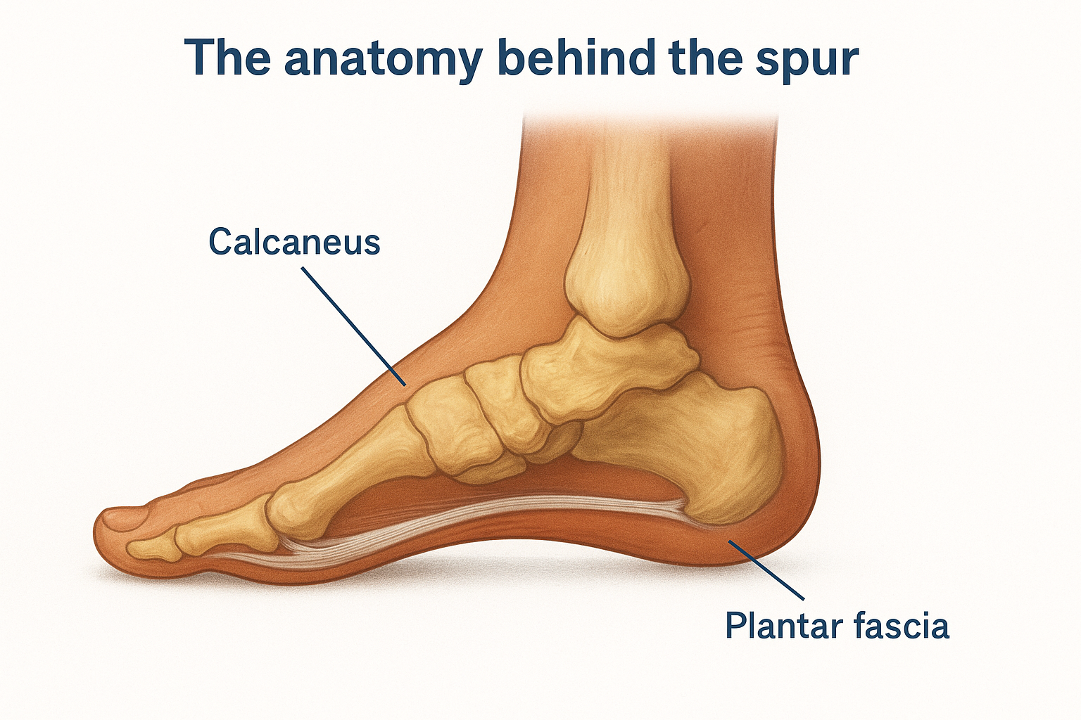
Location matters when it comes to symptoms. Spurs that grow along the inferior surface of the calcaneus, meaning the very bottom of the heel, are the most common and are most frequently linked to walking pain. Spurs that grow at the back of the heel where the Achilles tendon attaches are a separate condition called posterior heel spurs, and they tend to cause pain closer to the back of the ankle rather than the sole.
The connection to the plantar fascia
Running along the entire bottom of your foot is the plantar fascia, a thick band of connective tissue that links your heel bone to the base of your toes. This tissue supports your arch and absorbs shock during movement. When it is repeatedly strained, it pulls on the heel bone at its attachment point and triggers the calcium buildup that eventually becomes a spur.
This relationship is why heel spurs and plantar fasciitis so often appear together. Studies have found that a significant percentage of patients diagnosed with plantar fasciitis also show a heel spur on X-ray. However, having a spur does not automatically mean you have plantar fasciitis, and having plantar fasciitis does not mean you have a spur. The two conditions can exist independently or simultaneously, which is exactly why getting a proper diagnosis before starting any treatment matters so much.
Because the spur is rigid bone while the plantar fascia is flexible tissue, the interface between them becomes a focal point for irritation and inflammation with every step. First thing in the morning, the fascia has tightened overnight while you rest, so those first few steps stretch an already aggravated area, producing the sharp, stabbing sensation that most patients describe. Understanding this dynamic helps explain why treatment targeting only the visible spur on an X-ray often misses the bigger picture of what is actually causing your pain.
Why it happens and who is at risk
A bone spur on bottom of foot does not develop overnight. Your body creates one through a slow, defensive process that begins when the tissues attached to your heel bone experience repeated stress over a long period of time. Think of it as your skeleton trying to reinforce a weak point. Every time your heel absorbs impact and the plantar fascia pulls at its attachment site, tiny micro-injuries accumulate. Your body responds by laying down calcium at that stressed location, and after months or years, those deposits solidify into a visible bony growth.
The main mechanical causes
Foot mechanics play the largest role in why spurs form. When the arch of your foot is too flat (overpronation) or too high (supination), the force distribution across your heel becomes uneven, placing excessive tension on the plantar fascia and the heel bone itself. Walking, running, or standing for long periods amplifies that uneven stress with each repetition.
Footwear is another major driver. Shoes that lack adequate arch support or cushioning force the heel to absorb more impact than it should. Worn-out soles, thin-soled shoes, and high heels all contribute to the kind of repetitive strain that accelerates spur formation. Sudden changes in your activity level, such as starting a new exercise routine or increasing your mileage quickly, can also trigger the process before your foot has adapted.
The underlying mechanical problem, not just the spur itself, is what needs to be addressed for lasting relief.
Risk factors that raise your chances
Certain factors make some people more likely to develop a heel spur than others. Understanding where you fall on this spectrum helps you take preventive action earlier.
- Age: The plantar fascia loses elasticity as you get older, increasing the strain on the heel bone attachment.
- Body weight: Carrying excess weight puts significantly more load on your heels with every step.
- Occupation: Jobs that require standing or walking on hard surfaces all day create the sustained pressure that drives spur formation.
- Athletic activity: Runners, dancers, and athletes in high-impact sports face elevated risk due to repetitive heel loading.
- Tight calf muscles: Limited flexibility in the Achilles tendon and calf increases the pull on the plantar fascia.
Symptoms and how it differs from plantar fasciitis
Recognizing the symptoms of a bone spur on bottom of foot can feel confusing because the spur itself is rarely the direct source of pain. What you actually feel is the inflammatory response in the surrounding soft tissue, which is why symptoms can overlap heavily with other heel conditions. Knowing exactly what to look for helps you communicate more clearly with your podiatrist and avoid chasing the wrong diagnosis.
Common symptoms of a heel spur
The most frequently reported symptom is a sharp, stabbing pain on the underside of the heel that is worst during your first few steps in the morning or after sitting for a long period. Once you walk around and the tissue warms up, the pain typically eases to a dull ache. You may also notice tenderness when you press directly on the heel or discomfort that worsens after prolonged standing or physical activity.
Pain that improves with movement but returns after long periods of rest is one of the clearest signs that the plantar fascia and heel are involved.
Some people notice mild swelling or warmth at the base of the heel, though visible swelling is less common than with an acute injury. In more severe cases, the discomfort can spread into the arch of the foot.
How to tell it apart from plantar fasciitis
Plantar fasciitis and heel spurs share so many symptoms that they are easy to confuse, but the location and nature of the pain offer useful clues. With plantar fasciitis alone, pain tends to concentrate along the entire length of the arch and at the heel attachment point, and it is driven entirely by inflammation in the soft tissue band. A heel spur adds a structural component to that picture, meaning there is a physical bony growth present, but it does not necessarily make the symptoms more severe.
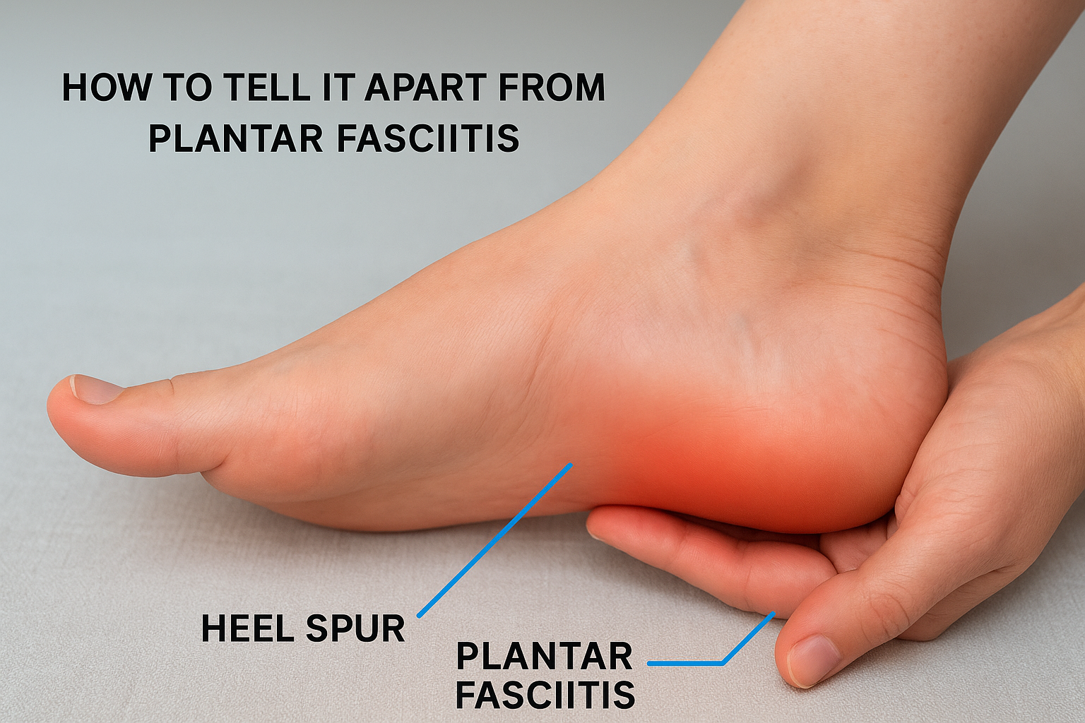
The key distinction is that plantar fasciitis is a soft tissue diagnosis while a heel spur is a structural finding on imaging. You can have significant plantar fasciitis pain with no spur visible at all, or you can show a clear spur on X-ray and feel minimal or no discomfort whatsoever. A clinical examination combined with imaging is the only reliable way to determine which condition, or combination of conditions, you are dealing with.
How doctors diagnose it
Diagnosing a bone spur on bottom of foot requires more than just looking at an X-ray. Your podiatrist needs to understand your full symptom history and movement patterns before ordering any imaging, because the spur showing up on a scan may not be the actual source of your pain. The diagnostic process is designed to build a complete picture so you get treatment targeting the right problem.
The physical examination
Your podiatrist will start by asking you to describe your pain: when it occurs, how long it has been present, and what activities make it better or worse. That history helps narrow down whether you are dealing with a structural issue, an inflammatory condition, or both at the same time.
From there, the physical exam involves pressing firmly along specific points on your heel and arch to pinpoint where the tenderness is concentrated. Your podiatrist will also assess your foot posture, range of motion in your ankle, and the flexibility of your calf muscles. Tight calf muscles and limited ankle dorsiflexion are common findings in patients with heel pain, and addressing them is often part of the treatment plan.
Where your pain is most intense on palpation tells your doctor far more than imaging alone can.
Imaging and what it reveals
A standard weight-bearing X-ray is the primary tool for confirming the presence of a heel spur. Because the X-ray is taken while you stand, it shows how the foot bears load and gives your podiatrist a more accurate look at the bony structures under real conditions. A spur appears as a visible projection extending from the calcaneus, and its size can be measured to track changes over time if needed.
Your podiatrist may also use diagnostic ultrasound to evaluate the soft tissue around the spur. Ultrasound shows the plantar fascia in real time and can reveal thickening, tearing, or active inflammation that an X-ray cannot capture. In cases where symptoms are severe or do not respond to initial treatment, an MRI may be ordered to rule out stress fractures, nerve entrapment, or other conditions that mimic heel spur pain. Getting the diagnosis right from the start is what allows your treatment plan to actually work.
At-home relief and self-care that helps
Managing a bone spur on bottom of foot at home is realistic for many people, especially in the early stages. The goal of self-care is to reduce inflammation in the surrounding soft tissue, improve the flexibility of the structures pulling on your heel, and take pressure off the aggravated area long enough for it to settle down. None of these measures remove the spur itself, but they can significantly reduce your day-to-day pain and functional limitations.
Stretching your plantar fascia and calf muscles
Stretching is one of the most effective things you can do at home. Because a tight plantar fascia and stiff calf muscles amplify the tension on your heel, loosening those structures reduces the load on the spur site directly. Before taking your first steps in the morning, sit on the edge of your bed and pull your toes back toward your shin for 30 seconds, holding the stretch through the arch of your foot. Repeat this two to three times before standing.
Stretching before your first step of the day is one of the simplest changes that produces a noticeable drop in morning pain.
Calf raises and seated towel stretches are also worth adding to your daily routine. Both target the Achilles tendon and soleus muscle, which run directly into the heel and contribute to the pulling force your plantar fascia has to resist.
Choosing the right footwear and insoles
Your shoes do more damage than most people realize. Thin-soled shoes, worn-out sneakers, and unsupportive flats increase the impact your heel absorbs with every step and keep the inflammation active. Switching to shoes with firm arch support, a slight heel cushion, and a sturdy midsole makes a measurable difference in daily discomfort.
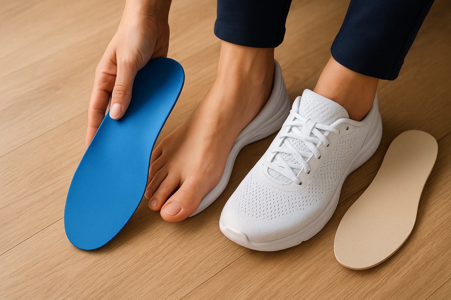
Adding over-the-counter heel cups or full-length orthotics inside your existing footwear can further cushion the spur site and redistribute pressure away from the most tender spot. Look for options with gel or foam cushioning specifically designed for heel pain.
Managing pain and swelling at home
Applying ice to the bottom of your heel for 15 to 20 minutes after activity helps control the inflammation that causes most of your pain. Avoid placing ice directly on skin; wrap it in a thin cloth. Anti-inflammatory medications like ibuprofen can also reduce short-term swelling and discomfort, though relying on them long-term without addressing the underlying mechanics is not a sustainable approach.
Treatments your podiatrist may recommend
When self-care does not provide enough relief, a podiatrist can offer a range of targeted interventions for a bone spur on bottom of foot that go well beyond what you can manage at home. The right treatment depends on how long you have had symptoms, the severity of your inflammation, and what your imaging shows. Most patients respond well to conservative professional care without ever needing surgery.
Custom orthotics and physical therapy
Your podiatrist may prescribe custom-molded orthotics that fit your exact foot shape and correct the mechanical issues driving the problem. Unlike over-the-counter insoles, custom orthotics are designed from a precise cast or scan of your foot, allowing them to redistribute pressure away from the spur site in a way generic products cannot replicate. Physical therapy is frequently prescribed alongside orthotics to address muscle imbalances and flexibility deficits that keep the plantar fascia under strain.
A physical therapist will guide you through targeted stretching protocols, strengthening exercises for the intrinsic foot muscles, and gait retraining if your walking pattern is contributing to the overload. These two interventions work together to attack the root mechanical cause, not just the symptoms.
Injections and advanced therapies
For patients with significant inflammation that has not responded to stretching and orthotics, corticosteroid injections deliver concentrated anti-inflammatory medication directly to the painful tissue around the spur. Your podiatrist will often use ultrasound guidance to place the injection precisely, improving both accuracy and effectiveness.
A single well-placed injection combined with ongoing mechanical correction often provides the turning point for patients who have struggled for months.
Extracorporeal shockwave therapy (ESWT) is another option your podiatrist may discuss. This non-invasive treatment uses focused sound wave energy to stimulate the body’s natural healing response in the chronically inflamed tissue surrounding the spur. Studies show it produces meaningful pain reduction in patients with stubborn heel pain who have not improved with other conservative measures.
Surgical options for persistent cases
Surgery is reserved for cases where 12 or more months of consistent conservative treatment have failed to provide adequate relief. The procedure typically involves releasing part of the plantar fascia and removing the spur through a minimally invasive approach, which reduces recovery time compared to traditional open surgery. Most patients return to normal activity within a few weeks following proper rehabilitation.
When to seek care and prevent it from coming back
Self-care goes a long way with a bone spur on bottom of foot, but there are clear situations where waiting longer makes the problem harder to treat. The earlier you get a professional evaluation, the more conservative your treatment options are likely to be. Delaying care until you can barely walk turns a manageable condition into one that may require more intensive intervention.
Signs you should not wait to see a podiatrist
Some symptoms point to a problem that home care cannot resolve on its own. If your heel pain has persisted for more than a few weeks despite stretching, better footwear, and rest, that is your signal to book an appointment. Pain that is spreading into the arch or up the back of the ankle, significant swelling that does not subside overnight, or numbness and tingling in the foot alongside heel pain all warrant prompt evaluation. These symptoms can indicate nerve involvement, stress fractures, or soft tissue tears that mimic a simple heel spur but need a different treatment approach entirely.
Waiting through months of worsening pain rarely improves your outcome and often extends the total recovery time.
You should also seek care if your pain is affecting how you walk. Compensating for heel pain by shifting weight onto the outer edge of your foot or your toes creates secondary problems in the knee, hip, and lower back that compound over time.
Steps to keep heel spurs from returning
Preventing recurrence starts with the same mechanics that caused the problem in the first place. Maintaining flexibility in your calf muscles and plantar fascia through consistent daily stretching keeps the tension on your heel bone manageable, even during periods of high activity. This is not a short-term fix; it is an ongoing habit that protects your heel long after the pain is gone.
Footwear choices matter just as much in prevention as they do during treatment. Replace worn-out athletic shoes before the midsole breaks down, and avoid walking barefoot on hard surfaces for extended periods. If your podiatrist prescribed custom orthotics, wear them consistently rather than only when your foot feels sore. Keeping your body weight within a healthy range also reduces the baseline load your heels carry with every step, lowering the chance that calcium deposits will accumulate again.
Getting back to comfortable walking
A bone spur on bottom of foot is a manageable condition for most people when you address both the structural spur and the soft tissue inflammation driving your symptoms. The steps covered in this article, from daily stretching and better footwear to professional orthotics and targeted therapies, work because they treat the root mechanical causes rather than just masking the pain. Most patients who follow through with a consistent treatment plan return to comfortable, unrestricted walking without needing surgery.
Your foot health affects everything you do, from your morning routine to your athletic performance. If heel pain has been slowing you down or getting worse despite your efforts at home, getting a professional evaluation is the most direct path forward. Schedule a same-day appointment at Achilles Foot and Ankle Center and get a clear diagnosis with a treatment plan built around your specific situation.